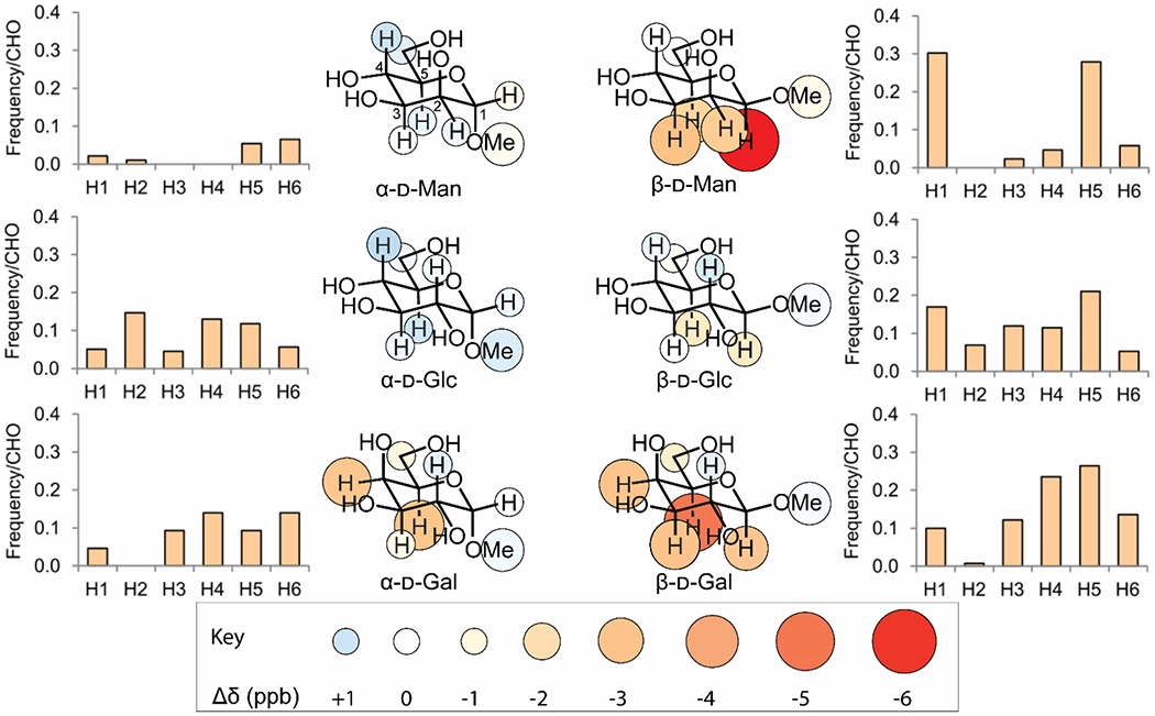
Figure 4. CH-π stacking in solution reflects patterns found in carbohydrate binding sites.

Inner panels show the change in chemical shift for each proton when 10 mM indole is added to a 0.5 mM solution of the indicated glycoside. Larger and darker red circles indicate stronger shielding from CH-π stacking. Outer panels show the probability of a binding site for the indicated monosaccharide having an aromatic group positioned closest to the indicated carbon. Data reproduced from Hudson, K. L.; Bartlett, G. J.; Diehl, R. C.; Agirre, J.; Gallagher, T.; Kiessling, L. L.; Woolfson, D. N., Carbohydrate-Aromatic Interactions in Proteins. J. Am. Chem. Soc. 2015, 137, 15152-15160. Copyright 2015 American Chemical Society. Figure adapted from Diehl 2021.95
