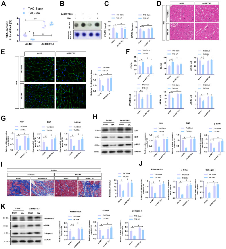
Figure 4.
The reverse effects of METTL3 over-expression on the anti-hypertrophy effect of MA in vivo. (A) The effect of METTL3 over-expression on the MA impaired total RNA m6A methylation content in TAC induced hypertrophic LV tissues detected with the EpiQuik m6A RNA Methylation assay kit and (B) confirmed by Dot Blot. (C) The ratio of HW/BW and HW/ TL shows the effect of METTL3 over-expression on the MA inhibited morphology of cardiac morphology. (D) Analysis of the effect of METTL3 over-expression on the MA preserved cardiomyocyte area in left ventricle through Histology H&E and (E) WGA staining of cardiac cross-sections. (F) Echocardiography detection of the parameters of LVEF, LVFS, LVEDV, LVESV, LVEDD, and LVESD to evaluate the effect of METTL3 over-expression on the MA preserved cardiac function of TAC mice. (G) Real-time PCR analysis of the mRNA levels of the hypertrophy markers (ANP, BNP, and β-MHC). (H) Western blot detection of the protein levels of the hypertrophy markers (ANP, BNP, and β-MHC). (I) Detection of the effect of MA on the TAC-induced myocardial fibrosis via Masson staining. (J) Real-time PCR analysis of the mRNA levels of the myocardial fibrosis (Fibronectin, Collagen I, and α-SMA). (K) Western blot analysis of the protein levels of the myocardial fibrosis (Fibronectin, Collagen I and α-SMA); *P<0.05, ** P <0.01 compared to the indicated group.

