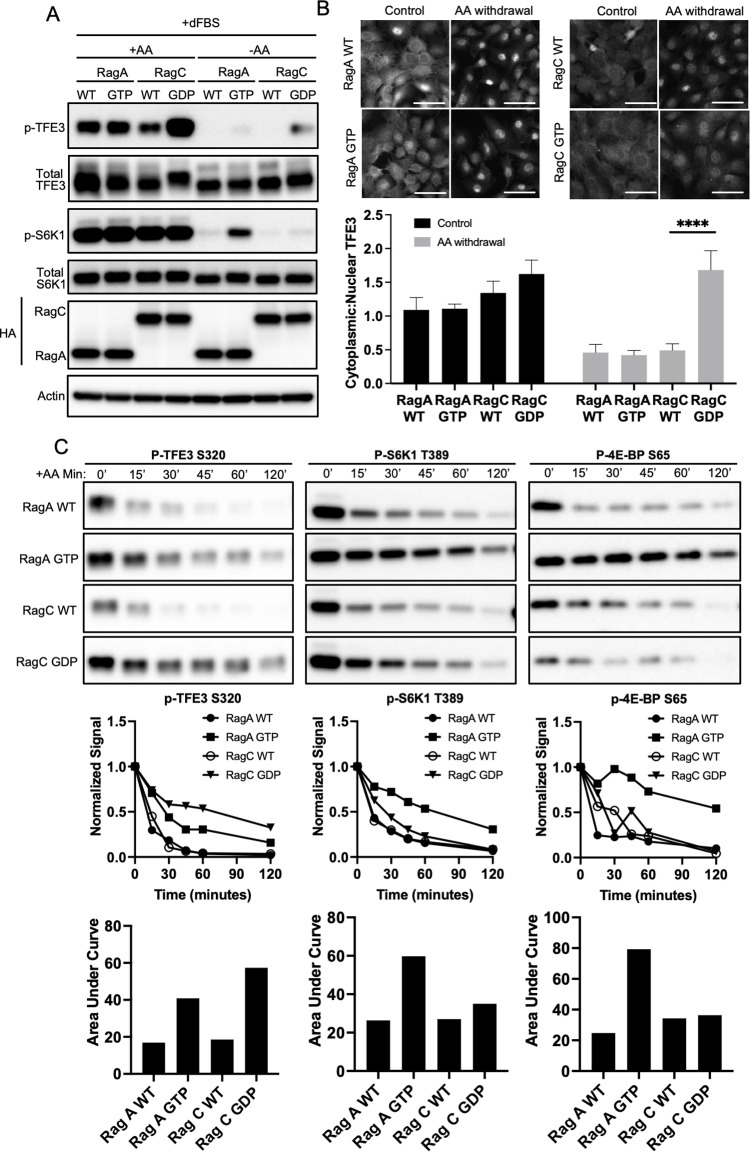
Fig 2. RagC, but not RagA, promotes TFE3 phosphorylation in response to AAs.
(A, B) C2C12 cells expressing HA-tagged WT or constitutive active RagA (GTP) or RagC (GDP) were switched from complete medium to media lacking AAs, as indicated, followed by immunoblotting for TFE3, phospho-TFE3, S6K, phospho-S6K, and HA (A) or immunohistochemistry for subcellular localization of TFE3 (B). Quantification of cytoplasmic-to-nuclear ratio of TFE3 is shown below the images. Scale bar: 20 μm, ****p < 0.0001 by Student t test. (C) The same cells as in A, subjected to a time course after withdrawal of AAs, followed by immunoblotting for phospho-TFE3, phospho-4EBP, and phospho-S6K. Densitometric quantification is shown below. The data underlying all the graphs shown in the figure is included in S1 Data. AA, amino acid; dFBS, dialyzed FBS; WT, wild type.

