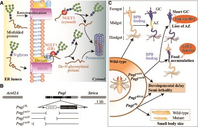
Fig. 1.

(A) Cartoon representation of retrotranslocation of misfolded N-glycosylated proteins from the ER lumen to the cytosol through a membrane-associated complex including HRD1, Derlin1 and VCP. These N-glycosylated proteins are deglycosylated either by cytosolic NGLY1 (top) or ER membrane-recruited NGLY1 (bottom) and thereafter undergo proteosomal degradation. (B) Schematic representation of the Pngl genomic region and different Pngl alleles generated so far. The dark area in Pngl represents the coding region. PnglPL is generated through CRISPR-Cas9 gene editing and harbours a nonsense mutation at codon 420 followed by the mini-white+ insertion. Pnglex14, Pnglex18 and Pnglex20 are microdeletions generated through imprecise excision of a P-element insertion in the Pngl locus. (C) Cartoon representation of some of the Pngl loss-of-function phenotypes and the impaired signalling pathways associated with these phenotypes. Not all alleles have been examined for all phenotypes. AZ, acid zone; GC, gastric caeca; BPB, bromophenol blue.
