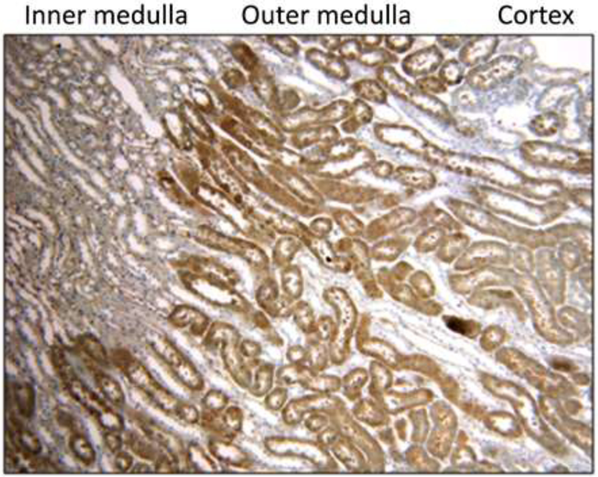
Figure 2:

Representative image of pimonidazole staining in the murine corticomedullary junction (100× magnification). Pimonidazole staining was primarily localized to the outer medulla. The staining pattern was similar between mice administered hyperoxia and normoxia.
