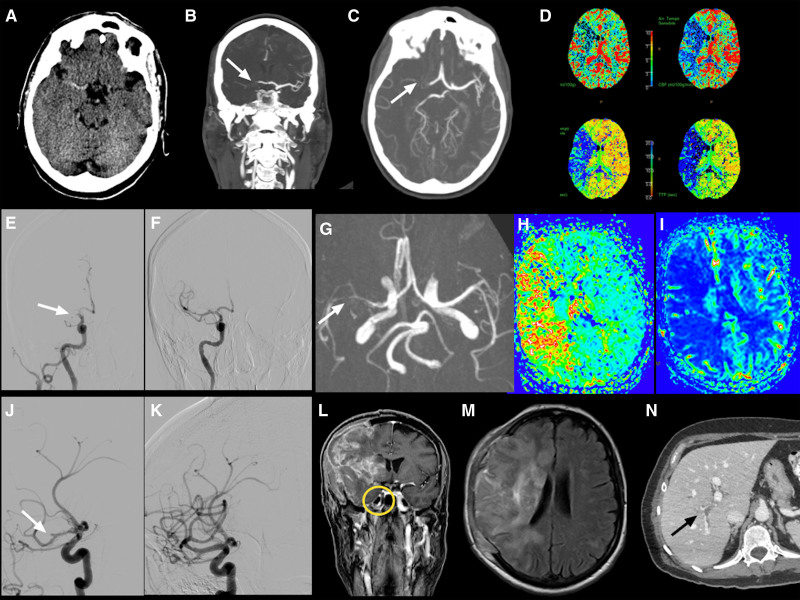
Figure 3.
Radiological findings from patient 6.
Case reported by De Michele et al.109 A, Computed tomography (CT) showed hyperdensity in the right middle cerebral artery. The CT angiography revealed proximal M1 segment occlusion of the right middle cerebral artery (MCA; white arrow in B and C); CT perfusion maps showed a large area of mismatch indicating salvageable penumbra (D); digital subtraction angiography confirming a proximal MCA occlusion (F) of the MCA occlusion (white arrow in E); MCA reocclusion on M2 segment 2 h after the procedure, 3-dimensional time-of-flight magnetic resonance imaging (MRI) sequence (white arrow in G), with extensive ischemic penumbra (time to peak map in H and cerebral blood flow in I). Second endovascular recanalization, oblique views showed occlusion of M2 segment (white arrow in J) with reopening of the vessel after the mechanical thrombectomy (K). Fourteen-day MRI follow-up after craniectomy showed the extension of ischemia to superficial and deep right MCA territory (M) with occlusion of right internal carotid artery at postcontrast sequences (yellow circle in L). N, Right portal vein thrombosis (black arrow).

