. 2022 Mar 21;11:e72555. doi: 10.7554/eLife.72555

Figure 8. Effects of R85G + R290 E, Y269D, Y279D, and F273D + R275 D mutations on the binding of LukE in live cells as determined by competition time-resolved fluorescence energy transfer (TR-FRET).

5 nM tracer ligands, CCL5-d2 for CCR5 (A) and ACKR1 (C) and CCL2-d2 for CCR2 (B), were used to determine TR-FRET at their respective receptors in the presence of LukE mutants or wild-type LukE. IC50 values were undetermined for LukE mutants due to loss of binding. Data shown is mean ± SEM of three independent experiments performed in triplicate.

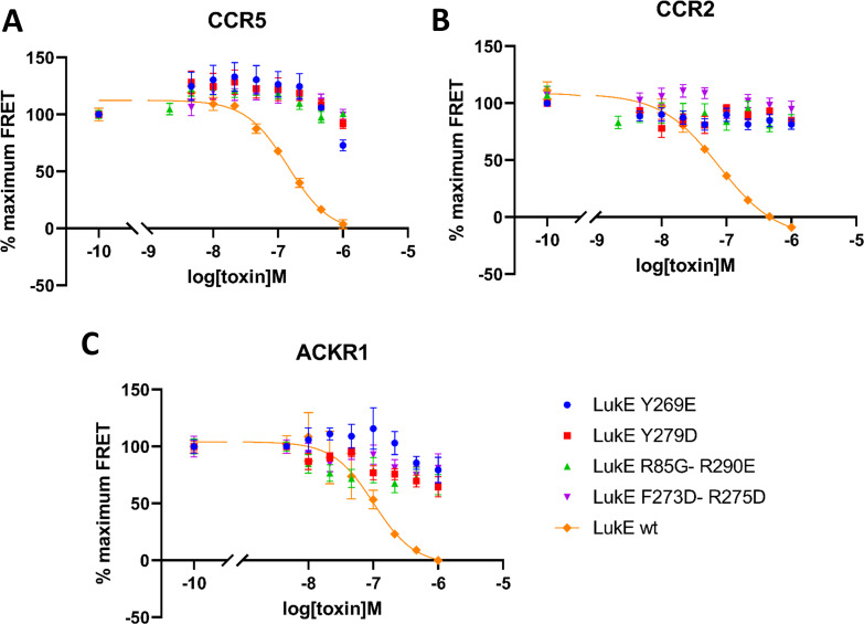
Figure 8.

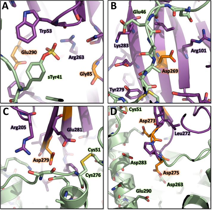
Figure 8—figure supplement 1. Environment of the mutated LukE residues in the ACKR1-LukE model.

Figure 8—figure supplement 1.

(A) Close up of sulfotyrosine binding site 1 in the ACKR1-LukE model with the R85G-R290E mutations. (B) Close up of sulfotyrosine binding site 2 in the ACKR1-LukE model with the Y269E mutation. (C) Close up of the orthosteric pocket near TM7 and extracellular loop 3 in the ACKR1-LukE model showing the Y279D mutation in the vicinity of the Cys51-Cys276 disulfide bridge. (D) Close up of the orthosteric pocket in the ACKR1-LukE model showing the F273D-R275D mutations.
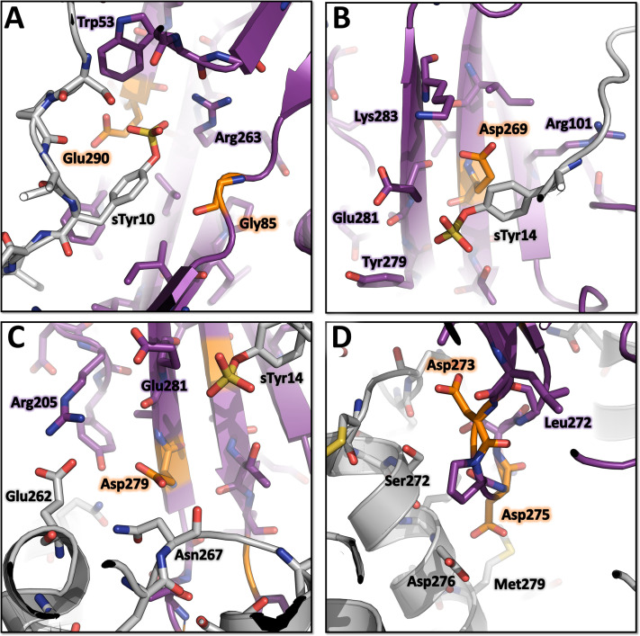
Figure 8—figure supplement 2. Environment of the mutated LukE residues in the CCR5-LukE model.

Figure 8—figure supplement 2.

(A) Close up of sulfotyrosine binding site 1 in the CCR5-LukE model with the R85G-R290E mutations. (B) Close up of sulfotyrosine binding site 2 in the CCR5-LukE model with the Y269E mutation. (C) Close up of the orthosteric pocket near extracellular loop 3 in the CCR5-LukE model showing the Y279D mutation. (D) Close up of the orthosteric pocket in the CCR5-LukE model showing the F273D-R275D mutations.