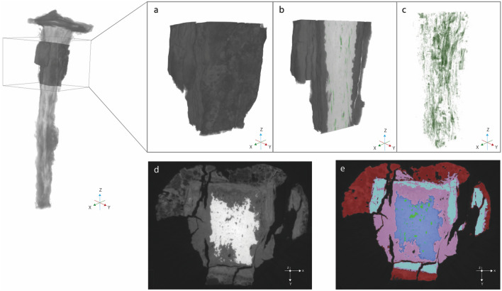
Figure 4.
High-resolution scans of a selected volume of the nails. (a) 3D rendering of analysed section (b) Sagittal cut of the 3D reconstruction, showing parallel structural discontinuities (c) 3D segmentation of structural discontinuities (green) (d) 2D slice, showing microstructure detail and corrosion stratigraphy in grayscale value (e) Segmented 2D slice in false colors: metal in blue, DPL in pink (goethite) and turquoise (magnetite) and TM (red).

