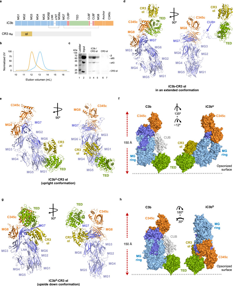
Fig. 1. Crystal structure of iC3b–CR3 αI.
a Schematic representation of the domain architecture of iC3b and the von Willebrand A (VWA) or inserted (I) domain of CR3 (CR3 αI). b Analytical SEC (anaSEC) traces of iC3b–CR3 αI (orange) and iC3b (blue). c Coomassie-stained SDS-PAGE electrophoresis of peak fractions for the iC3b–CR3 αI chromatogram shown in (b). We repeated at least five independent purifications of the iC3b–CR3 αI complex with similar results. d Ribbon representation of the extended conformation of the iC3b–CR3 αI complex. e Ribbon representation of the iC3bU–CR3 αI complex shown in two orientations related by a 90° rotation around a vertical axis on the plane of the figure. f Comparison of C3b (left) with iC3bU (upright conformation) with respect to a hypothetical opsonized surface covalently anchored to the TED domain via Gln991. C3b and iC3bU are shown as molecular surfaces, and the CUB domain of C3b is shown in white. The orientation of the TED domain in C3b and iC3b is the same. The rotations necessary to superimpose C3b into iC3bU are indicated. g Ribbon structure of the iC3bD–CR3 αI complex. h As in g for iC3bD (upside-down conformation). The same color scheme is applied to all panels: the β-chain is shown in light blue, the α65 chain in slate blue (except the TED domain, which is shown in green), the α43 chain in orange, and the CR3 αI domain in olive. Complexes in panels (d–f) are shown in the same orientation for clarity’s sake. Source data are provided as a Source Data file.

