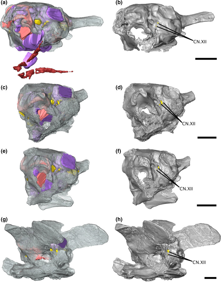
FIGURE 4.

Isosurface renderings of the varanopid braincase in left posterolateral view, showing the relative positioning of the hypoglossal foramina. (a‐b) Mesenosaurus, (c‐d) ROMVP87043, (e‐f) ROMVP86543, (g‐h) Aerosaurus. Scale bars equal 5 mm. CN.XII, hypoglossal nerve
