
FIGURE 3.
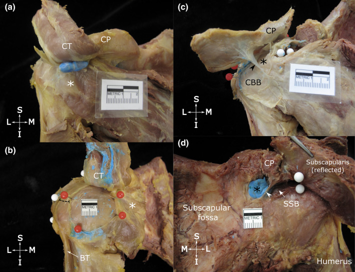
Location and dimensions of the coracobrachial (a and b) and subcoracoid bursa (c) and the relationship between the superior extension of the subtendinous bursa of subscapularis and the subcoracoid bursa (d). Anterior view of the shoulder showing (a) the typical location of the coracobrachial bursa (unopened, injected with blue latex), deep to the tip of the coracoid process (CP), conjoint tendon (CT, reflected superiorly) and overlying the subscapularis tendon (white asterisk); and (b) a large, circular coracobrachial bursa (opened, latex removed) with its floor exposed, delineated with red pins. The distance between the anterior boundary of the subacromial bursa (white pins) and lateral border of the coracobrachial bursa is indicated by the dashed line. (c) shows an opened subcoracoid bursa (black asterisk) that extends deep to the coracoid process (CP, resected and reflected superiorly) and shares a wall (arrowhead) with a coracobrachial bursa (CBB). (d) shows a subcoracoid bursa (filled with blue latex) and superior extension of the subtendinous bursa of subscapularis (SSB, arrow), separated by a thin wall (arrowhead). The attachment of the subtendinous bursa of subscapularis to the deep surface of the subscapularis tendon is indicated by white pins. Abbreviations: BT, biceps tendon (long head); I, inferior; L, lateral; M, medial; S, superior. Scale bar marked in cm