
FIGURE 4.
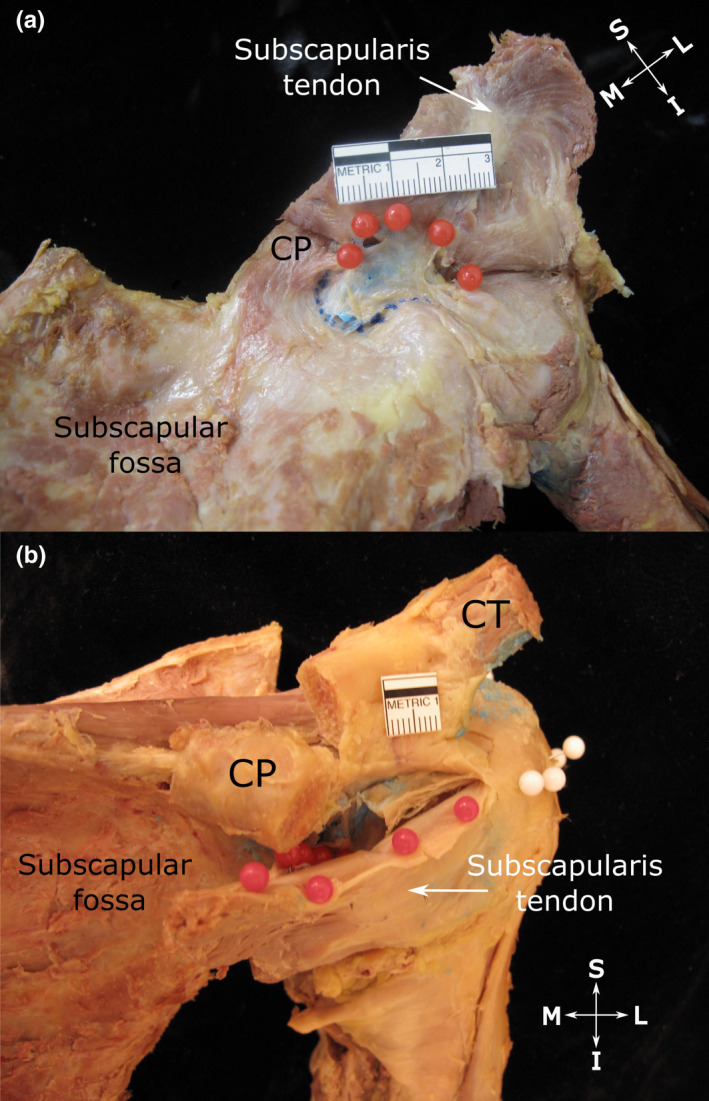
Subtendinous bursa of subscapularis (a) and extent of its superior extension along the anterior surface of the subscapularis tendon (b). (a) Anterior view of the shoulder showing the subtendinous bursa of subscapularis (injected with blue latex). Its roof (indicated by red pins) attaches to the upper aspect of the deep surface of the subscapularis tendon and its floor (indicated by blue dotted line) fuses with the neck of the scapula and root of the coracoid process (CP, tip resected) as well as the anterior glenohumeral joint capsule. (b) Anterosuperior view of the shoulder showing the subtendinous bursa of subscapularis, overhanging the upper edge of the subscapularis tendon and attaching to its superficial surface (indicated by red pins). The close relationship between the upper edge of the subscapularis tendon along with its bursal attachment to the underside of the coracoid process (CP, tip resected and reflected) is also visible. White pins indicate the extent of the subacromial bursa, but are not relevant to this image. Abbreviations: I, inferior; L, lateral; M, medial; S, superior. Scale bar marked in cm