
FIGURE 6.
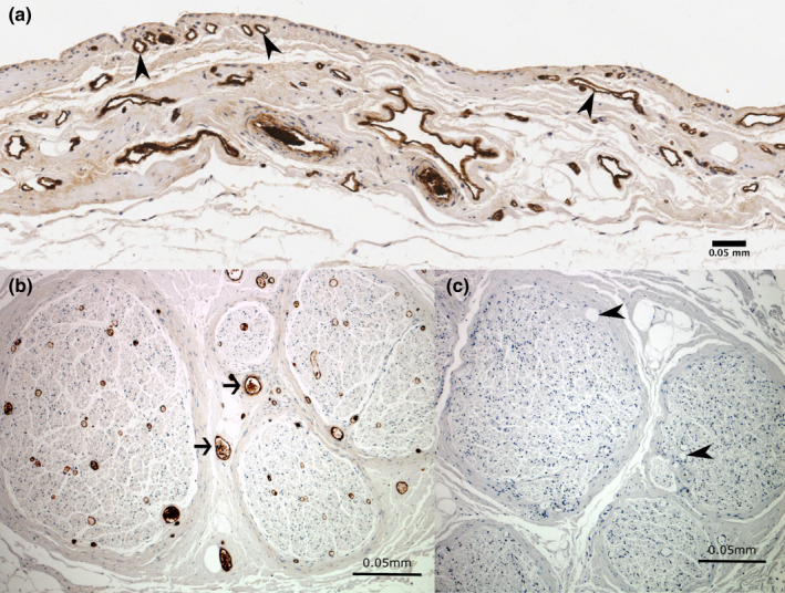
Immunoreactivity for von Willebrand factor showing blood vessels in the (a) subacromial bursa and (b) positive control tissue. Sections showing immunoreactivity to von Willebrand factor in (a) blood vessels in the subacromial bursa (arrowheads) and (b) blood vessels (arrows) in the positive control section within the sciatic nerve. The negative control section (c) shows no immunoreactivity to von Willebrand factor in blood vessels (arrowheads) or other non‐specific staining. Cell nuclei are stained blue (haematoxylin)