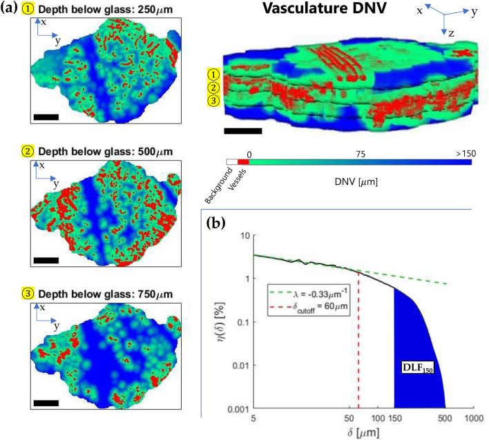
Figure 2.
Evaluation of the diffusion-limited fraction and convexity index λ: (a) 2D visualizations of the colour-coded distance-to-nearest-vessel (DNV) map at three different depths below the window chamber to tissue interface, from the application of the 3D Euclidean distance transform to the binarized microvasculature within the tumour VOI. The upper right of the figure shows the full 3D vasculature and tissue distances parametric volumetric image; (b) The corresponding log–log DNV histogram for the full tumour VOI. The indicated and metrics quantify short and long-distance properties of the DNV, respectively. Scale bars are mm, 3D volumes are mm3 (). Each distance bin was μm, the rescaled isotropic voxel size in the 3D parametric images for smooth computations.

