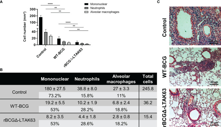
Figure 7.
Characterization of the cellular infiltrate in the lungs. Total number of mononuclear cells, neutrophils, and alveolar macrophages in the perivascular and peribronchiolar infiltrates in the lungs of BALB/c mice infected only (Control), or immunized and challenged (BCG and rBCGΔ-LTAK63) represented as cell counts per mm². **p < 0.01 and ****p < 0.0001 (A) and the actual cell numbers, percentage and total cell count in each group (B). Five images at 40 x magnification per lung lobule, totaling 25 images per treatment, were randomly selected. For cell counting, ImageJ software and the Color Deconvolution 2 plugin were used to visualize and separate nuclei from the cytoplasm. Representative histopathology of lungs sections stained with H&E (bar, 20 µm) (C). Challenge experiments were performed twice.

