Figure 5.
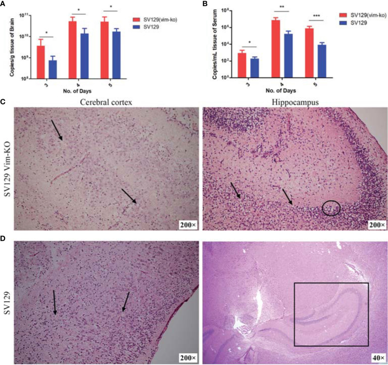
Detection of viral load in serum and brain and description of brain histopathological damage after infection of SV129 and SV129 (Vim-KO) mice with dengue virus (DENV)-2. (A, B) Changes in the viral load in the brain and serum; *P < 0.05; **P < 0.01; ***P < 0.001. (C, D) Brain histopathological section on the 5th day after infection in SV129 and SV129 (Vim-KO) mice. (C) Left: the cortical stratifications disappeared, with a large number of apoptotic pyknotic cells (black arrow). Right: the hippocampus displayed apoptotic pyknosis of glial cells (black arrow) and local cerebral liquefactive necrosis (black circled). (D) Left: the cortical stratification was normal, and apoptotic pyknosis of glial cells (black arrow) was found in all six cortical layers. Right: the hippocampus (black square) showed no obvious abnormalities.

