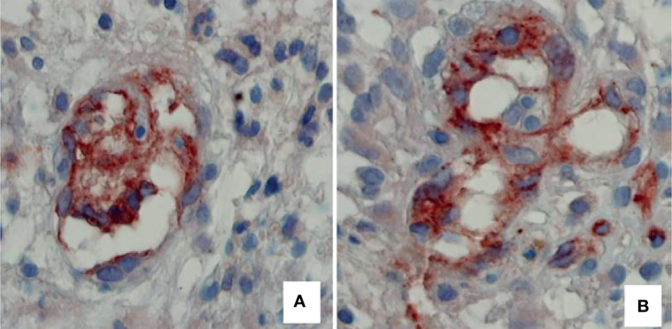
Figure 3.

Two examples of tumor vessels, respectively, with a low and high number of connections of intraluminal tissue folds with the opposite vascular wall, expression of intussusceptive microvascular growth in II malignancy grade tumor specimen (A), compared with IV malignancy grade (B). Blood vessels have been identified by immunohistochemical reaction with an anti-CD31 antibody. Original magnification: x 60 (Reproduced from 4).
