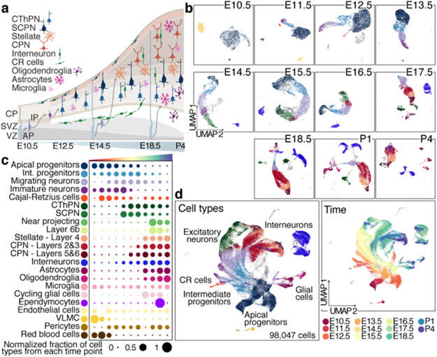
Figure 1. Comprehensive atlas of murine cortical development.
a Cellular diversity and development of the neocortex.
b UMAP visualization of scRNA-seq data from Individual time points. Cells are colored by cell type assignement.
c Normalized contribution of each time point to each cell type present in the developing cortex. See also Extended Data Fig. 1.
d Combined time points visualized by age (left), or cell types (right), legend in c.
VZ: ventricular zone, SVZ: subventricular zone, CP: cortical plate, CR: Cajal-Retzius cells, AP: apical progenitors, IP: intermediate progenitors, CThPN: corticothalamic projection neurons, SCPN: subcerebal projection neurons, CPN: callosal projection neurons.

