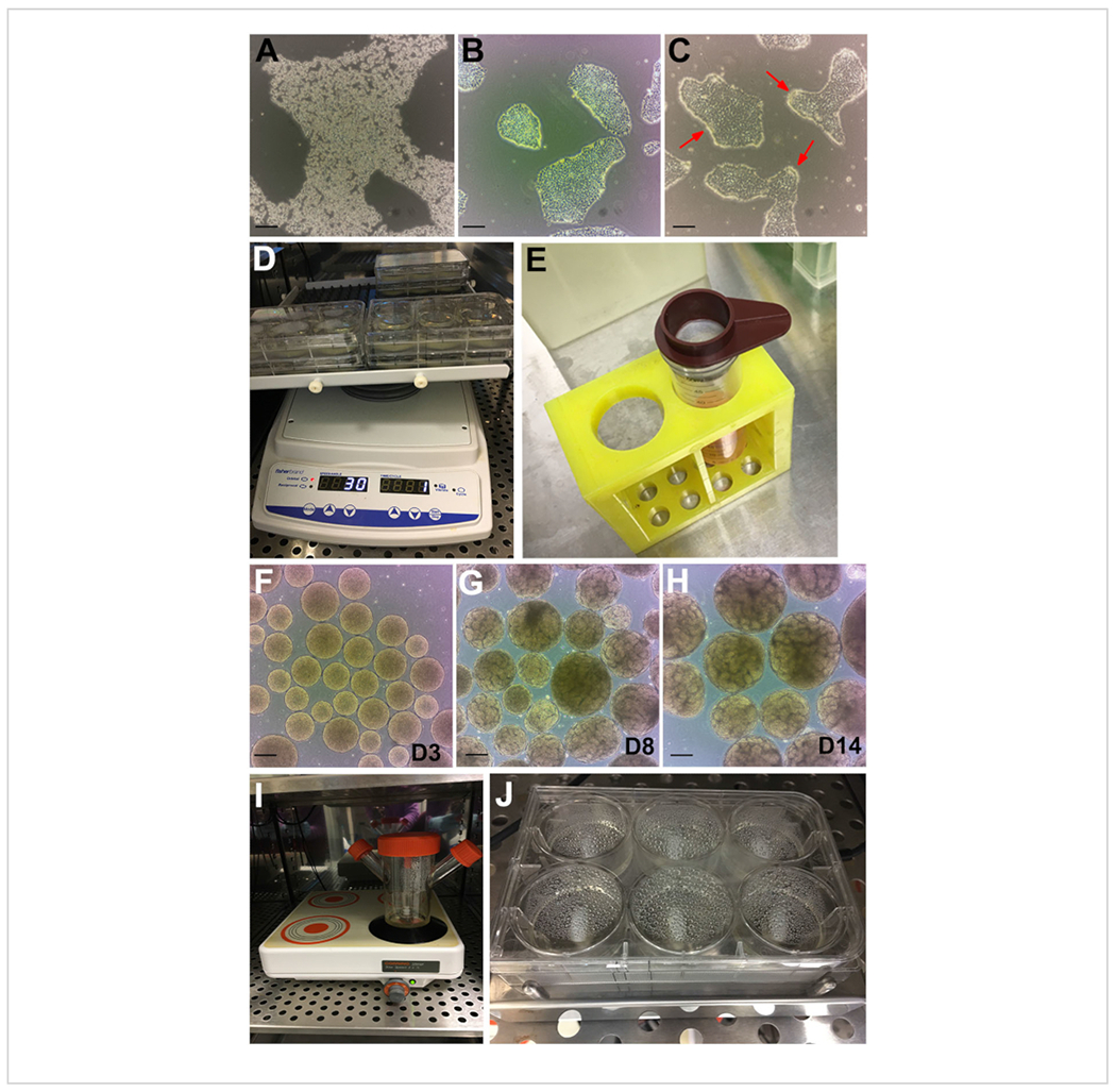
Figure 2: Stages of the protocol.

(A) Bright-field image of hPSC colony treated with GCDR. (B) Optimal confluency, and colony size to begin a kidney organoid assay. (C) hPSCs treated with dispase for 6 minutes. Red arrows point to edges of the colonies curling up. (D) Organoid assays on an orbital shaker. (E) Use of 200 μm cell strainer to sieve out large embryoid bodies. (F) Embryoid bodies at day 3 (D3) before transferring to Stage II medium. (G) Emergence of tubule formation can be observed at day 8 (D8) and (H) optimal timepoint for organoid harvesting and treatment at day 14 (D14). (I) Spinner flask used for bulk culture on a multi-position magnetic plate. (J) Assay on a multi-well magnetic stir plate. Scale bars, 200 μm. Please click here to view a larger version of this figure.
