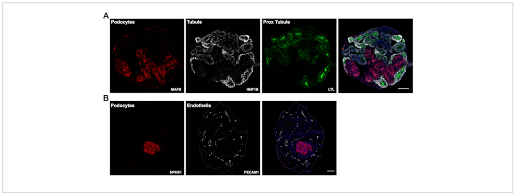
Figure 3: Expected results.

(A) Representative confocal images of immunofluorescently labeled paraffin sections of day 14 kidney organoids showing positive staining for tubule epithelia (HNF1B and LTL) and podocyte clusters (MAFB). (B) Day 26 kidney organoid sections labeled for podocyte clusters (NPHS1) and endothelial cells (PECAM1). Scale bars, 100 μm (A); 200 μm (B). Please click here to view a larger version of this figure.
