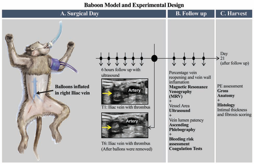
Figure 1: Baboon model of iliac vein venous thrombosis and experimental design.
A. This model used a 6 hour temporary balloon occlusion in juvenile baboons. On surgical day 0, standard contrast venography and ultrasound was performed in order to acquire baseline information regarding the vein to be thrombosed. A thrombus is created in the iliac vein by threading one balloon catheter via the internal jugular vein caudally down the inferior vena cava by fluoroscopy, to just below the iliac bifurcation. Another catheter was moved cranially up the femoral vein near the pelvic crest. This isolated a vein segment of approximately 3.0 cm in length. During the 6 hours of venous occlusion, the area between balloons were monitored hourly for absence of flow and increasing echogenicity representative of thrombus formation, using ultrasonography. After 6 hours, the balloon catheters were removed, incisions closed, an ultrasound performed to confirm the presence of a thrombus within the iliac vein and the animal monitored for post-operative recovery.
B. Animals were evaluated on Days 2, 7, 14, and 21; hematology and coagulation tests, venous duplex ultrasound imaging, magnetic resonance venography (MRV), and standard contrast venography.
C. On Day 21, the animals were humanely euthanized and vein samples harvested from the thrombosed iliac (experimental samples) and from the non-thrombosed iliac (control samples) for histology.

