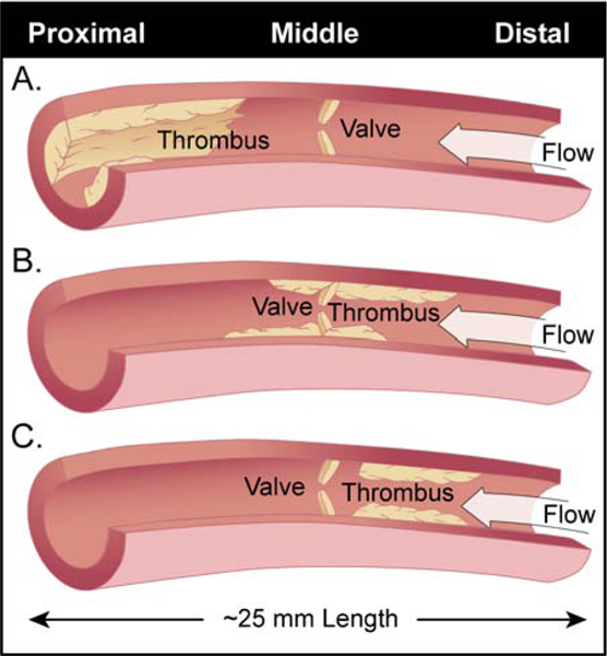
Figure 6: Clinical scenarios that can affect iliac valve reflux times in this study.
All animals were evaluated for thrombus burden and valve reflux times via duplex ultrasound imaging. The study area of the right iliac vein consisted of a proximal (Prox.), middle (Mid.) and distal (Dist.) segments for each animal which all together made up an iliac vein segment approximately 25 mm in length.
Panel A. Control animal number 5 had a large thrombus burden in the proximal iliac segment that did not incorporate the valve yielding a viable reflux time with a small flow channel present. These animals had decrease lumen patency and increased vein wall inflammation.
Panel B. Control animals 3 and 4 had non-occlusive middle iliac segment thrombus that incorporated the iliac valve through day 14 post thrombosis. However, by day 21, these animals had chronic anterior and posterior thrombus present with competent valves via duplex ultrasound. Treated animals 3 and 4 had non-occlusive middle iliac thrombus that incorporated the iliac valve through day 7 and 14 post thrombosis respectively. However, by day 21, these animals had complete resolution of thrombus burden but non-competent valves via duplex ultrasound.
Panel C. Control animals 1 and 2; Treated animals 1 and 2 had venous thrombi in the distal iliac segment that did not incorporate the iliac valve. The treated animals however had lower thrombus burden than the controls animals leading to increased lumen patency and decreased vein wall inflammation.
Note: Secondary venous valves above and/or below interrogated segment and/or technical error should be considered.

