FIGURE 8.
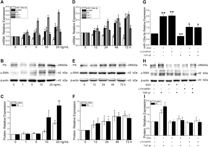
TGF-β1 reduces miR-130a-3p expression via Smad3 signaling pathways. MRC-5 cells were stimulated with various concentrations of TGF-β1 for 48 h or with 5 ng/ml of TGF-β1 for different periods of time. (A,D) qRT-PCR quantitation of mRNA expressions of miR-130a-3p and fibrosis-related genes. (B-C,E-F) Representative and statistical data of α-SMA and FN proteins. *p < 0.05, **p < 0.01, ***p < 0.001, versus 0 ng/ml or 0 h, by the Mann–Whitney U test, n = 3–5. MRC-5 cells were pretreated with 10 μM of TGF-βRII inhibitor (LY2109761), Smad3 inhibitor (SIS3), or control (DMSO) for 30 min and then stimulated with 10 ng/ml of TGF-β1 for 24 h. (G) qRT-PCR quantitation of mRNA expression of miR-130a-3p. **p < 0.01, versus the control group; & p < 0.05, versus the SISI3 group; # p < 0.05, versus the LY2109761 group, by the Mann–Whitney U test, n = 4–6. (H–I) Representative and statistical data of α-SMA and FN proteins. *p < 0.05, **p < 0.01, versus the control group; & p < 0.05, versus the TGF-β1 group, by the Mann–Whitney U test, n = 4-5.

