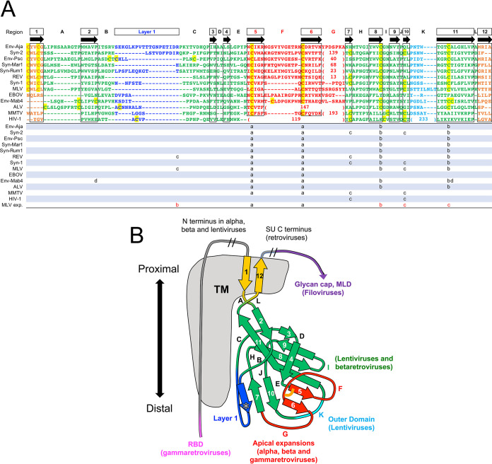
FIG 5.
Conserved orthoretroviral and filoviral PDs. (A) Orthoretroviral and filoviral PD sequences structurally aligned to the PD of Env-Aja. Note that the sequences are not aligned by sequence similarity but rather represent residue alignments in three-dimensional space to show the equivalent β-strand regions of the PD structure and the boundaries of connecting loops. The structural alignments were generated by the DALI server, except for the terminal β-strands 1 and 12. These two β-strands, which are not part of the core PD and are modeled in different orientations in different models, cannot be automatically aligned and were aligned manually. Insertions relative to the minimal Env-Aja SU model are not shown to highlight the β-strands shared by the models and structures, except for insertions in the major variable loop F and K regions, which are shown by the number of amino acid residues in parentheses. The lengths of all main loop regions within the PD are shown at the bottom of Fig. 7. Only one representative of each orthoretroviral and filoviral lineage is shown. The consensus β-strands are indicated by boxes and arrows. Cysteine residues are highlighted in yellow. Disulfide bonds in models and structures are shown below the structural alignment in matching lowercase letters, with the last line indicating the experimentally determined disulfide bonds in the MLV SU. The proximal domain is highlighted in green, except for the apical, layer 1, and loop K regions, which are highlighted in red, blue, and cyan. Terminal β-strands 1 and 12 are highlighted in orange. The boundaries of the HIV-1 gp120 and EBOV GP1 sequences correspond to residues labeled as aa 31 to 497 of chain A and aa 62 to 187 of chain I in the structures under PDB accession numbers 3JWD and 3CSY, respectively. (B) Schematic representation of the PD structures and major extended regions in different viral lineages. Regions are colored as described above for panel A, with the PD, apical region, layer 1, and K regions shown in green, red, blue, and cyan. The location of HIV-1 gp41 relative to the PD in the trimer structure is shown in gray. The asterisk in layer 1 indicates a β-strand that sometimes extends the β-sheet formed by extended β-strands 7 and 10. The conserved disulfide bond between β-strands 5 and 6 in the apical domain is shown as an orange line. The regions with major expansions in different lineages are indicated. MLD, mucin-like domain.

