
FIG 6.
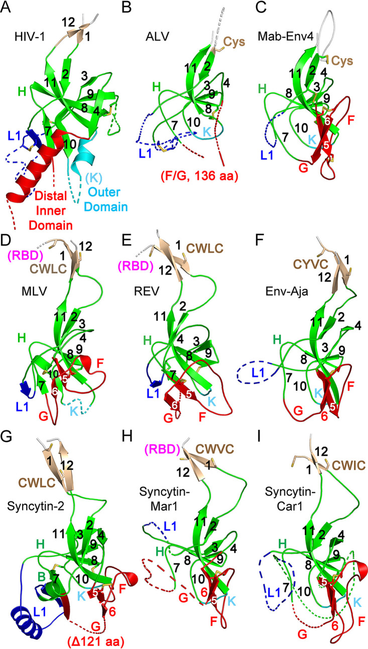
Conserved orthoretroviral SU proximal domain β-sandwich. (A) Structure of the HIV-1 gp120 PD region (PDB accession number 3JWD). (B to I) PD regions of the ALV (B), Mab-Env4 (C), MLV (D), REV (E), Env-Aja (F), syncytin-2 (G), syncytin-Mar1 (H), and syncytin-Car1 (I) SU models. Parts of layer 1 (L1) and regions F and G and the HIV-1 gp120 distal inner and outer domains are shown as dotted lines for clarity. The PD, apical domain, and layer 1 regions are shown in green, red, and blue. Selected loop and β-strand regions are labeled in each panel. Cysteine residues are shown as sticks. The locations of the conserved CWLC consensus motifs of gammaretroviral SU in β-strand 1 are indicated in panels D to I. The connections to the RBD in the models in panels D, E, and H are shown in magenta. All structures and models are shown in the same orientation as those in Fig. 2 and 3.