FIG 4.
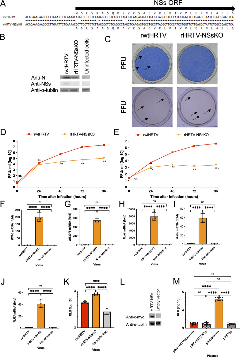
An analysis of the function of HRTV NSs in type I interferon-mediated innate immunity. (A) Alignment of the genomic sense S RNA coding sequences of rwtHRTV and rHRTV-NSsKO. The methionine at the translation initiation codon of the NSs protein was substituted with alanine, and two stop codons were inserted into the NSs protein ORF. (B) Vero 9013 cells were infected at an MOI of 0.1 FFU/cell with recombinant viruses. On 3 days postinfection (dpi), cell lysates were immunoblotted to detect the indicated proteins. (C) Shown is a comparison of plaque and focus morphologies of rwtHRTV and rHRTV-NSsKO. Plaques and focuses are pointed out by arrows. Shown are comparisons of replication kinetics of rwtHRTV and rHRTV-NSsKO in Vero 9013 cells (D) and A549 cells (E). Shown are the IFN-β (F), human ISG15 (G), MxA (H), RIG-I (I), and TLR3 (J) mRNA levels in A549 cells infected with each recombinant virus. (K) Activation levels of the ISRE promoter in 293T cells infected with each recombinant virus at an MOI of 2. 293T cells were transfected with pISRE-TA-luc or pTA-luc and pRL-TK. First, the cells were incubated for 15 h at 37°C and then inoculated with recombinant viruses. After 48 h, the activities of Fluc and Rluc were measured. (L) Expression of recombinant NSs proteins in 293T cells. (M) Activation levels of the ISRE promoter in 293T cells, which transiently expressed recombinant NSs protein and were stimulated with IFN-β. 293T cells were transfected with pISRE-TA-luc or pTA-luc, pRL-TK, and pKS-HRTV-NSs-c-myc or pKS336 (empty vector cells). First, the cells were incubated for 48 h and then treated with IFN-β. After 18 h, the activities of Fluc and Rluc were measured. Error bars represent the SD. ****, P < 0.0001; ***, P < 0.001; ns, P > 0.05. Experiments shown in D and E were performed in triplicate wells for each condition and repeated three times. Experiments shown in F, G, H, I, and J were performed three times. Experiments shown in K and M were performed in quadruplicate and sextuplicate, respectively.

