FIGURE 2.

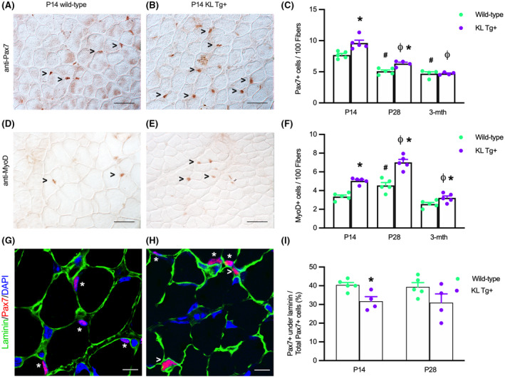
KL Tg expression increases numbers of satellite cells and activated myoblasts during early postnatal development (A, B) Representative images of Wt (A) and KL Tg+ (B) quadriceps muscle at P14 immunolabeled for Pax7 (reddish‐brown nuclei). (C) Numbers of Pax7+ cells per 100 fibers in quadriceps from Wt and KL Tg+ at P14, P28, and 3 months mice. (D, E) Representative images of Wt (D) and KL Tg+ (E) quadriceps muscle at P14 immunolabeled for MyoD (reddish‐brown nuclei). (F) Numbers of MyoD+ cells per 100 fibers in quadriceps from Wt and KL Tg+ at P14, P28, and 3 months mice. For A, B, D, and E, open arrowheads (>) indicate Pax7+ (A, B) or MyoD+ (D, E) labeled cells. Bar = 50 μm. For C and F, *indicates significantly different from age‐matched Wt control at p < .05 analyzed by t‐test. #Indicates significantly different from P14 Wt at p < .05 analyzed by one‐way ANOVA with Tukey's multiple comparisons test. ϕIndicates significantly different from P14 KL Tg+ at p < .05 analyzed by one‐way ANOVA with Tukey's multiple comparisons test. Error bar represents SEM. N = 4 or 5. (G, H) Representative images of Wt (G) and KL Tg+ (H) quadriceps muscle at P14 immunolabeled for Pax7 (red), laminin (green), and DNA labeled with DAPI (blue). *Indicates Pax7+ cells under the basal lamina. Open arrowheads (>) indicate Pax7+ cells outside of laminin. Bars = 10 μm. (I) Ratio of Pax7+ cells under the basal lamina to total Pax7+ cells in sections of quadriceps muscles from P14 and P28 Wt and KL Tg+ mice. *Indicates significantly different from age‐matched Wt control at p < .05 analyzed by t‐test. Error bar represents SEM. N = 4 or 5
