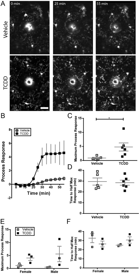
Figure 7.
(A) Representative images of microglial response to focal laser ablation insult over the course of 1 hour in binocular primary visual cortex of Cx3Cr1G/+ mice exposed to either vehicle or TCDD during gestation and lactation. Notice the microglial processes accumulating at the autofluorescent injury core over the course of the experiment. Scale bar = 20μm. (B) Time course of microglial process response to focal laser ablation insult in mice exposed to vehicle or TCDD. (C) Mice exposed to TCDD exhibit a significantly higher microglial process response to focal laser ablation injury than vehicle-exposed mice (n = 5 animals per group; unpaired t-test, P = 0.0384). (D) There is no significant difference in speed of process response across groups. *P < 0.05; Graphs show individual data points and mean ± SEM.

