. 2022 Mar 9;11:e76963. doi: 10.7554/eLife.76963

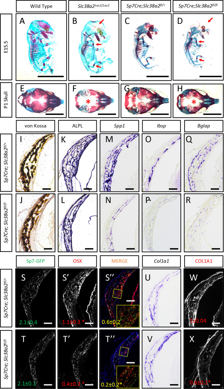
Figure 5. Slc38a2-dependent proline uptake is required for osteoblast differentiation during bone development.

(A–H) Skeletal preparations of Slc38a2LacZ/LacZ or wildtype controls (A, B, E, F) or Sp7Cre;Slc38a2fl/fl or Sp7Cre;Slc38a2fl/+ littermate controls (C, D, G, H) at embryonic day (E)15.5 (A–D) or P1 (E–H). Red arrow (A–D) or asterix (E–H) highlights reduced mineralization. A total of n = 7 or n = 5 Slc38a2LacZ/LacZ animals and n = 5 or n = 5 for Sp7Cre;Slc38a2fl/fl animals were analyzed at E15.5 or postnatal day (P)1, respectively. Scale bar = 5 mm. (I–R) Representative von Kossa staining (I, J), alkaline phosphatase (ALPL) staining (K, L) in situ hybridization for Spp1 (M, N), Ibsp (O, P), Bglap (Q, R), and Col1a1 (U, V), or immunofluorescent staining for OSX (S’, S”, T’, T”) and COL1A1 (W, X) on Sp7Cre;Slc38a2fl/fl (n = 4) (J, L, N, P, R, T, V, X) or Sp7Cre;Slc38a2fl/+ (n = 4) (I, K, M, O, Q, S, U, W) newborn calvariae. *p≤0.05 by paired two-tailed Student’s t-test. Scale bar = 100 μm.

Figure 5.

Figure 5—figure supplement 1. Slc38a2 is required for osteoblast differentiation in vitro.
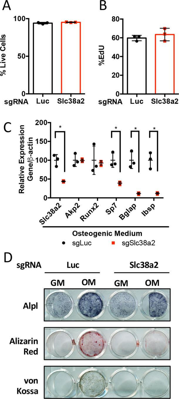
Figure 5—figure supplement 1.

(A–D) Effect of Slc38a2 targeting on cell viability (n = 3) (A), EdU incorporation (n = 3) (B), mRNA expression (n = 3) by qPCR analysis (C), or functional assays (D) in calvarial cells cultured in growth media (GM) or osteogenic medium (OM) for seven or 10 days (n = 3). Error bars depict SD.
*p≤0.05 by unpaired two-tailed Student’s t-test. See numerical source data in Figure 5—figure supplement 1—source data 1.
Figure 5—figure supplement 1—source data 1. Numerical source data for Figure 5—figure supplement 1.
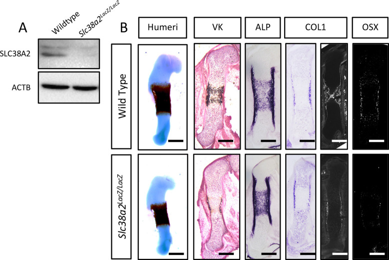
Figure 5—figure supplement 2. Slc38a2LacZ/LacZ mutants have impaired osteoblast differentiation during endochondral ossification.

Figure 5—figure supplement 2.

(A) Western blot analysis of SNAT2 expression in femur bone shaft protein isolated from Slc38a2LacZ/LacZ or wildtype controls. (B) Representative images of humerus skeletal preparations, von Kossa staining, alkaline phosphatase (ALPL) staining, in situ hybridization for Col1a1, or immunofluorescence staining for OSX and COL1A1 on femur sections of embryonic day (E)15.5 Slc38a2LacZ/LacZ or wildtype controls littermate controls (n = 3 animals). Scale bar = 200 μm. See uncropped Western blot images in Figure 5—figure supplement 2—source data 1.
Figure 5—figure supplement 2—source data 1. Uncropped western blot source data for Figure 5—figure supplement 2.
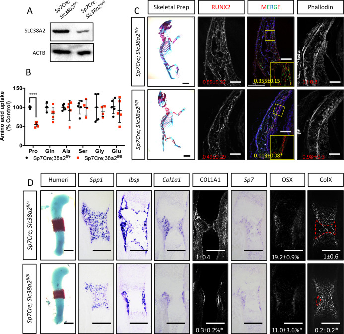
Figure 5—figure supplement 3. Sp7Cre;Slc38a2fl/fl have impaired osteoblast differentiation during endochondral ossification.

Figure 5—figure supplement 3.

(A) Western blot analysis of SNAT2 expression in femur bone shaft protein isolated from Sp7Cre;Slc38a2fl/fl or Sp7Cre;Slc38a2fl/+ littermate controls (n = 3). (B) Evaluation of amino acid uptake in femurs isolated from newborn Sp7Cre;Slc38a2fl/fl or Sp7Cre;Slc38a2fl/+ littermate controls (n = 5). (C) Skeletal preparations of newborn Sp7Cre;Slc38a2fl/fl or Sp7Cre;Slc38a2fl/+ littermate controls (n = 5). Scale bar in skeletal prep = 5 mm. Phalloidin staining or immunofluorescence staining for RUNX2 on postnatal day (P)0 Sp7Cre;Slc38a2fl/fl or Sp7Cre;Slc38a2fl/+ calvariae (n = 4). Scale bar = 100 μm. Boxed region shown in inset image. (D) Representative images of in situ hybridization for Spp1, Ibsp, Col1a1, and Sp7, or immunofluorescence staining for OSX, COL1A1, and COLX on humerus sections from embryonic day (E)15.5 Sp7Cre;Slc38a2fl/fl or Sp7Cre;Slc38a2fl/+ littermate controls (n = 4 animals). Red dashed line denotes the remodeling area. Fold change ± SD. Error bar depicts SD. *p≤0.05, ****p≤0.00005 by paired two-tailed Student’s t-test. Scale bar = 200 μm. See numerical source data and uncropped Western blot images in Figure 5—figure supplement 3—source data 1.
Figure 5—figure supplement 3—source data 1. Numerical and uncropped western blot source data for Figure 5—figure supplement 3.