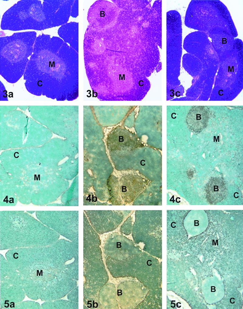
FIG. 3-5.
Fig. 3. Histopathologic evaluation of thymic tissue. (a) Thymic tissue representative of saline-treated uninfected and ZDV-treated uninfected groups. (b and c) Thymic tissue representative of the saline-treated FIV-infected (b) and ZDV-treated uninfected (c) groups. C, cortex; M, medulla; B, B-cell follicle. Magnification, ×33. Fig. 4. Immunohistochemical analysis of thymic tissue stained for B-cell CD21 expression. (a) Thymic tissue representative of the saline-treated uninfected and ZDV-treated uninfected groups. (b and c) Thymic tissue representative of the saline-treated FIV-infected (b) and ZDV-treated FIV-infected (c) groups. C, cortex; M, medulla; B, B-cell follicle. Magnification, ×33. Fig. 5. Immunohistochemical analysis of thymic tissue stained for cytokeratin expression. (a) Thymic tissue representative of the saline-treated uninfected and ZDV-treated uninfected groups. (b and c) Thymic tissue representative of the saline-treated FIV-infected and ZDV-treated FIV-infected groups. C, cortex; M, medulla; B, B-cell follicle. Magnification, ×33.

