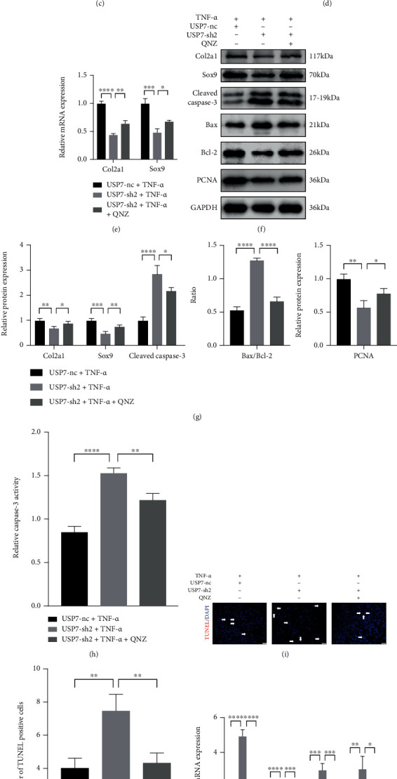
Figure 7.

NF-κB signaling inhibitor QNZ reverses chondrocyte proliferation, apoptosis, and inflammatory response caused by USP7 knockdown under TNF-α-induced inflammation. (a) Relative BiP and CHOP mRNA expression in USP7 knockdown and its control groups under TNF-α-induced inflammation after 48 h chondrogenic induction, with and without QNZ. (b) p-eIF2α, eIF2α, ATF4, CHOP, p-p65, and p65 protein expression of in USP7 knockdown and its control groups under TNF-α-induced inflammation after 48 h chondrogenic induction, with and without QNZ. (c) Quantitative measurement of B. (d) Alcian blue and toluidine blue staining in USP7 knockdown and its control groups under TNF-α-induced inflammation after 48 h chondrogenic induction, with and without QNZ. Scale bars = 100 μm. (e) Relative Col2a1 and Sox9 mRNA expression in USP7 knockdown and its control groups under TNF-α-induced inflammation after 48 h chondrogenic induction, with and without QNZ. (f) Col2a1, Sox9, Cleaved Caspase-3, Bax, Bcl-2, and PCNA protein expression of in USP7 knockdown and its control groups under TNF-α-induced inflammation after 48 h chondrogenic induction, with and without QNZ. (g) Quantitative measurement of F. (h) Relative Caspase-3 activity in USP7 knockdown and its control groups under TNF-α-induced inflammation after 48 h chondrogenic induction, with and without QNZ. (i) TUNEL staining in USP7 knockdown and its control groups under TNF-α-induced inflammation after 48 h chondrogenic induction, with and without QNZ. White arrows indicated TUNEL-positive cells. Scale bars = 50 μm. (j) Quantitative measurement of (i). (k) Relative IL-6, COX, NOS2, and MMP13 mRNA expression in USP7 knockdown and its control groups under TNF-α-induced inflammation after 48 h chondrogenic induction, with and without QNZ. (l) IL-6 expression in USP7 knockdown and its control group supernatant under TNF-α-induced inflammation after 48 h chondrogenic induction, with and without QNZ. ∗p < 0.05, ∗∗p < 0.01, ∗∗∗p < 0.001, and ∗∗∗∗p < 0.0001.
