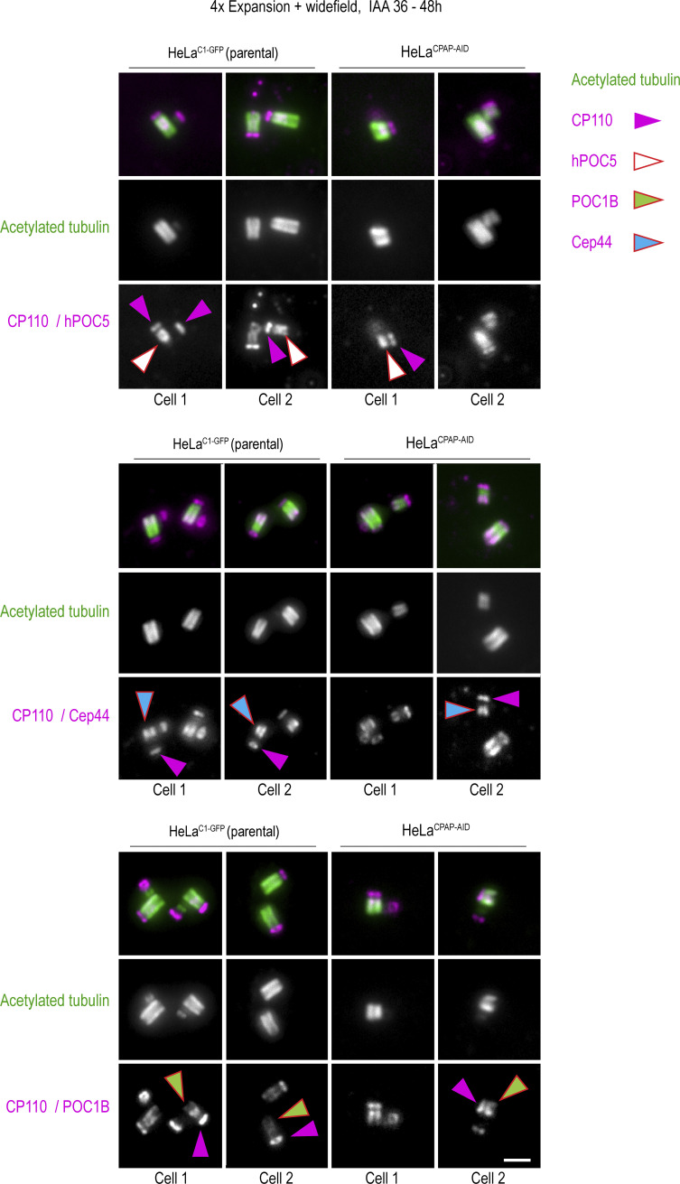
Figure S5.
Localization of centrosomal proteins on expanded HeLaC1-GFP and HeLaCPAP-AID centrioles after 36–48 h of IAA treatment. Cells were treated with IAA for 36–48 h, fixed, expanded, and immunolabeled for indicated centriolar proteins (magenta) and acetylated tubulin (green). Both control centrioles and short centrioles contain indicated centrosomal proteins. Arrows point to individual proteins, to facilitate interpretation. Proximal-distal polarity is preserved on short and narrow centrioles in IAA-treated cells. On short centrioles, proximal and distal portions are both shorter than normal. Scale bar: 2 µm (corresponding to ∼0.25 µm in unexpanded sample). This figure is associated with Fig. 6 of the main text.

