Figure S3.
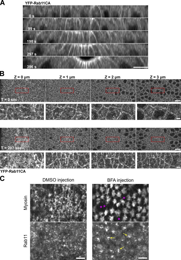
Rab11 vesicles are likely involved in the exocytic pathways. (A and B) Constitutively active Rab11 localizes to the plasma membrane in addition to vesicles and perinuclear compartments. (A) Montage showing the cross-section view of an embryo expressing YFP-Rab11CA during early ventral furrow formation. T = 0 s indicates the onset of apical constriction. Apical and lateral membrane localization of Rab11CA is enhanced over time in the constricting cells. N = 4 embryos. Scale bar, 10 μm. (B) En face view of the same embryo at T = 0 and 297 s from the apical surface (0 μm) to 4 μm below the surface. Scale bar, 10 μm. Bottom panels for each time point shows enlarged view of regions indicated by red boxes. Scale bar, 2 μm. (C) Acute inhibition of secretory pathway by Brefeldin A injection results in enlarged Rab11 structures (yellow arrows) compared to control DMSO injection. Breaks between neighboring myosin (magenta asterisks) was often observed in BFA injected embryos. N = 3 embryos. Scale bars, 5 μm.

