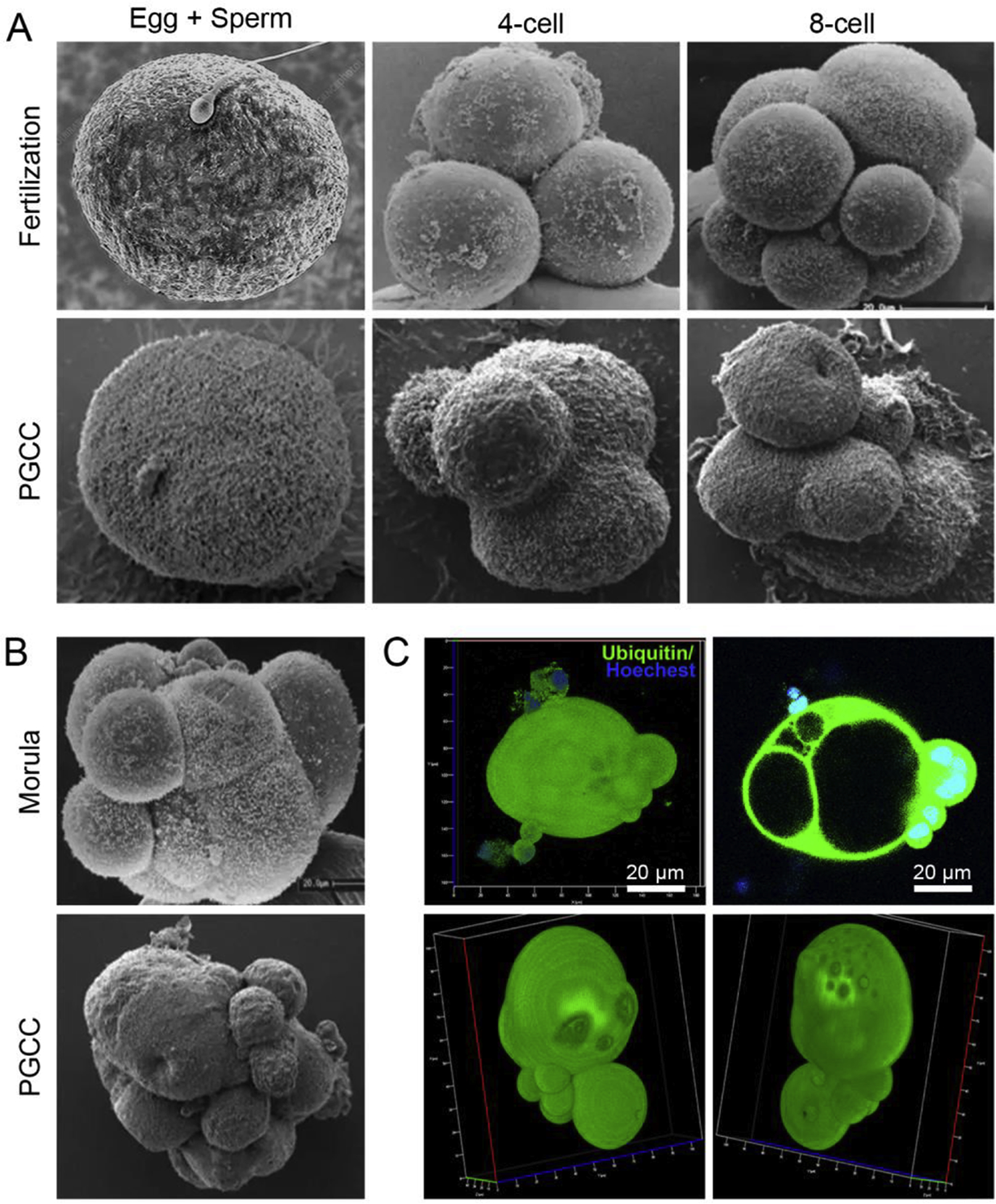
Fig. 2. Growth and division of polyploid giant cancer cells (PGCCs) mimics that observed in blastomere-stage embryos.

A. Stages of cleaved blastomeres following fertilization. The lower panels of A and B showing the morphology of PGCCs mimicking blastomeres were visualized by scanning electron microscopy and are adapted from our previous publication [54]. B. Scanning confocal microscopy images showing PGCCs with a blastocyst-like structure as compared with human morulae. C. 3D confocal scanning images of Hey-derived PGCC (upper panel) and a spheroid derived from Hey-derived PGCC (low panel). The zygote image is adapted from https://www.sciencephoto.com/media/873684/view/human-egg-and-sperm-sem (credit, Dennis Kunkel, microscope/science photo library with permission). The 4-cell, 8-cell, and morula stage embryo images are adapted from a figure previously published in Biology of Reproduction [163], reproduced with permission from Dr. George Nikas and the journal. The images of PGCCs were adapted from our previous publication [54].
