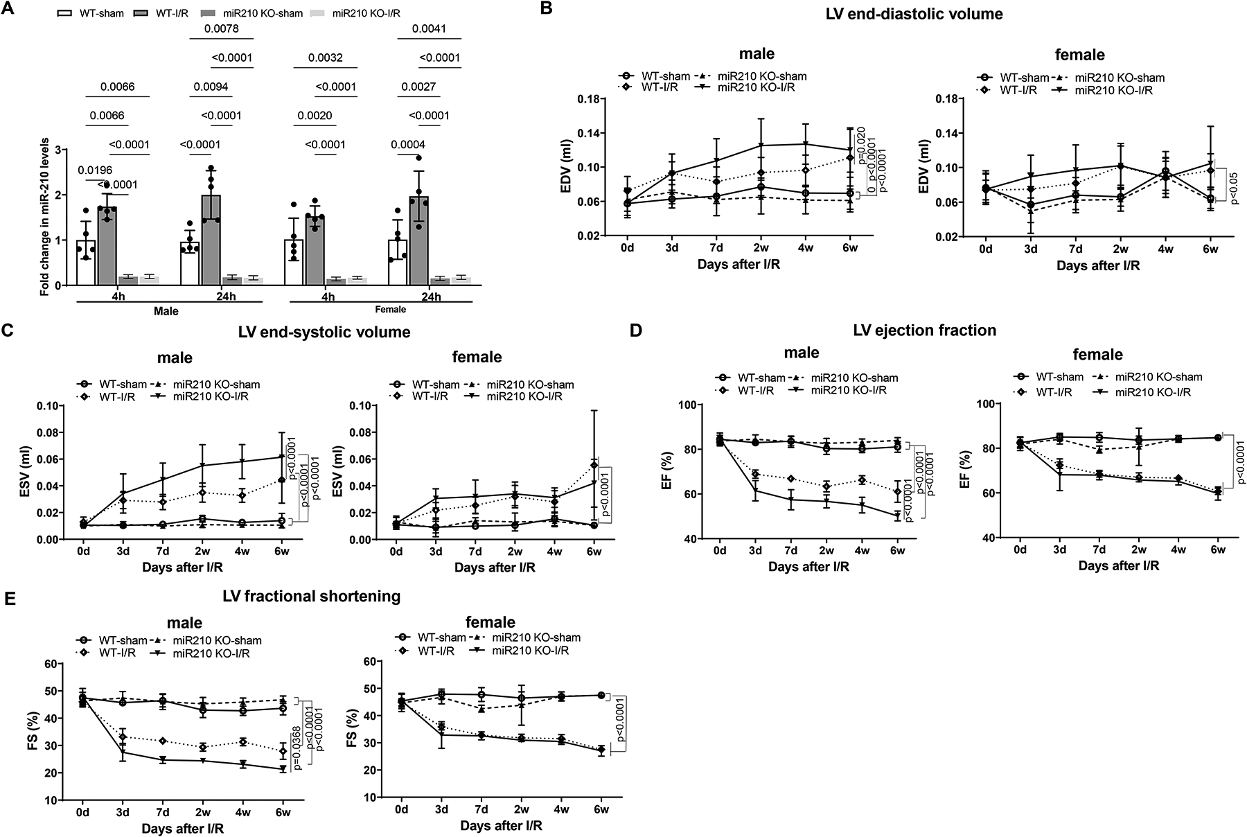
Figure 1. MiR-210 deficiency exaggerates cardiac dysfunction after myocardial infarction in male mice.

MiR-210 knockout (KO) and their wild-type (WT) littermate control mice with C57Bl/6 background at 2 months old underwent myocardial infarction (MI) induced by in vivo heart ischemia and reperfusion (IR) treatment via ligation of the mid-left anterior descending coronary artery for 30 min followed by reperfusion. Sham-operated animals without ligation of LAD coronary artery served as naïve controls. Hearts were isolated after 4 h or 24 h reperfusion and miR-210 in the heart was measured (A). Cardiac function was evaluated by echocardiography prior to or up to 6 weeks after in vivo IR treatment (B-E). Data are means ± SD, with n=5–6 animals per group, and analyzed by three-way ANOVA (A) or two-way repeated-measures ANOVA (B-E) followed by Tukey’s test. P values are shown in the Figure.
