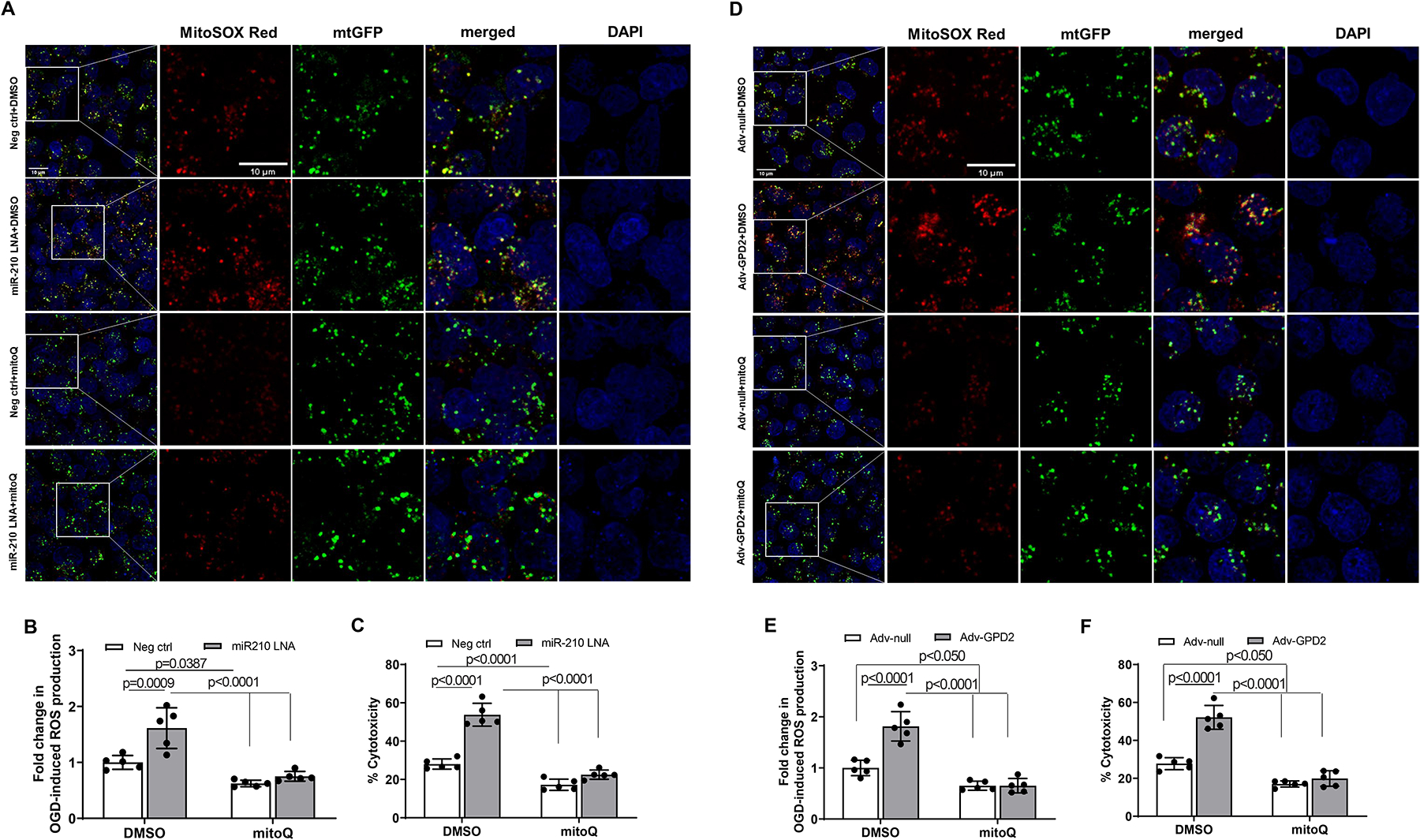
Figure 7. MitoQ inhibits miR-210 LNA and GPD2 overexpression-induced mtROS generation and cardiomyocyte hypoxic injury.

Mouse neonatal cardiomyocytes were treated with miR-210 LNA (50 nM) or scramble LNA (Neg. Ctrl), Adv-GPD2 (1.6 × 10⁷ pfu/ml) or Adv-null in the presence of MitoQ (250 nM) or vehicle control DMSO overnight, followed by subjecting to 2 h OGD and 24 h reoxygenation. Mitochondria-derived ROS was visualized with MitoSOX Red and fluorescence was colocalized with mitochondria, as visualized with Mitochondria-GFP (mtGFP) using fluorescent confocal microscopy (A and D). Panels B and E show the quantitative data of mtROS. Cell injury was measured with LDH release assay (C and F). Data are means ± SD with n=5 independent cultures per group, and analyzed by two-way ANOVA followed by Tukey’s test. P values are shown in the Figure.
