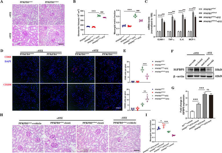
Fig. 6. A mutation in PFKFB3 blocks the expression of proinflammatory molecules and the migration of macrophages in the kidneys of diabetic mice.
A Representative images of PAS-stained kidneys. Scale bar, 50 μm. B Quantification of glomerular volume and mesangial matrix fraction per mouse. n = 5–6 mice. C RT–PCR analysis of the mRNA levels of ICAM-1, TNF-α, IL-6, and MCP-1 in control and diabetic mice. n = 5–6 mice. D Representative images of CD68 and CD206 immunofluorescence. Scale bar, 50 μm. E Quantification of CD68- and CD206-positive cells in diabetic and non-diabetic PFKFB3wt/mut or PFKFB3wt/wt mice. F, G Western blot analysis and densitometric quantification of IGFBP5 protein levels in the kidney lysates of PFKFB3wt/wt + vehicle, diabetic PFKFB3wt/wt + vehicle, diabetic PFKFB3wt/wt + lentivirus, and diabetic PFKFB3wt/mut + lentivirus mice. n = 5 mice. H Representative images of PAS-stained kidneys. Scale bar, 50 μm. I Quantification of the mesangial matrix fraction per mouse. n = 5 mice. The data are shown as the mean ± SD. *P < 0.05, **P < 0.01, ***P < 0.001; ###P < 0.001; ns, no significance.

