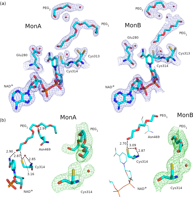
Fig. 5. Cofactor and substrate structures in the ALDH1A3-NAD+ complex (PDB code 7QK8).
a 2Fo–Fc electron density maps contoured at σ = 1 over Cys313, Cys314, Glu280, NAD+, PEG1, PEG2 and water molecules inside the cofactor and substrate-binding pockets. b For monomers A and B, hydrogen bonds of Cys314, Asn469 and NAD+ with PEG1, and polder OMIT maps at σ = 3.5 over Cys314 and PEG1. A highly conserved water molecule near Cys314 is depicted in red. The parts of the structure depicted with thin lines in monomer B represent those no longer interacting with the PEG1 molecule, with respect to monomer A.

