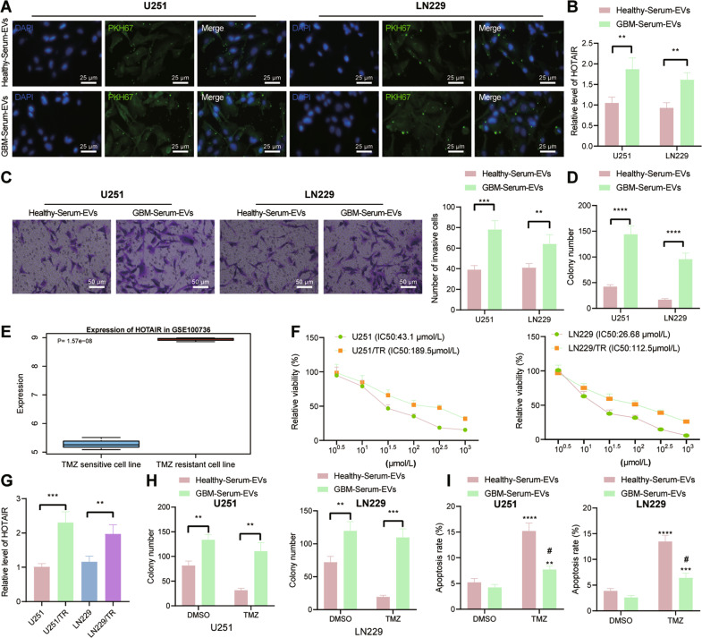
Fig. 2. GBM-serum-EVs induce the malignant features as well as TMZ resistance of GBM cells by mediating HOTAIR.
A Representative images of internalization of serum-EVs by U251 and LN229 cells under a fluorescence microscope. B RT-qPCR detection of HOTAIR expression in U251 and LN229 cells co-cultured with GBM-serum-EVs and healthy-serum-EVs. **p < 0.01. C Invasion of U251 and LN229 cells co-cultured with GBM-serum-EVs and healthy-serum-EVs measured by Transwell assay. **p < 0.01, ***p < 0.001. D Colony formation of U251 and LN229 cells co-cultured with GBM-serum-EVs and healthy-serum-EVs measured by colony formation assay. ****p < 0.0001. E HOTAIR expression in the GSE100736 data set. F IC50 of U251, U251/TR, LN229, and LN229/TR cells co-cultured with GBM-serum-EVs and healthy-serum-EVs measured by CCK-8. G RT-qPCR detection of HOTAIR expression in TMZ-resistant U251/TR and LN229/TR cells and its parental cells U251 and LN229. **p < 0.01, ***p < 0.001. H Colony formation of U251/TR and LN229/TR cells are co-cultured with GBM-serum-EVs and healthy-serum-EVs measured by colony formation assay. **p < 0.01, ***p < 0.001. I Flow cytometric analysis of apoptosis of U251/TR and LN229/TR cells co-cultured with GBM-serum-EVs and healthy-serum-EVs. ****p < 0.0001, compared with cells treated with DMSO. #p < 0.05, compared with healthy-serum-EVs. Each experiment was repeated three times independently.

