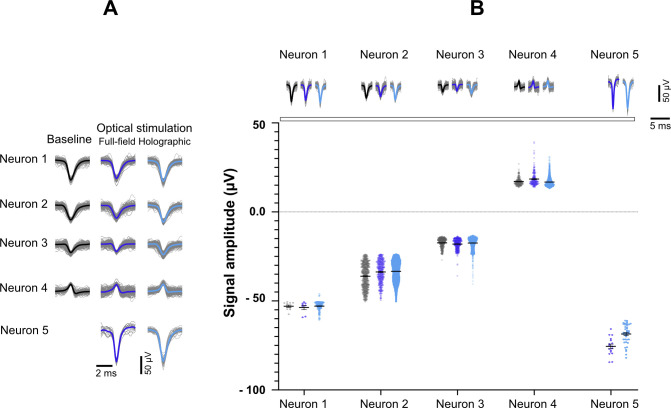
Figure S3. Recorded signals of active neurons detected in a single electrode at baseline, full-field, and holographic stimulation.
(A) Each waveform represents the activity recorded from one neuron under different conditions. Neuron number 5 was only active during full-field and holographic stimulation. (B) Comparison of AP amplitudes recorded from each neuron under the three conditions. There was no significant difference between waveform amplitude at full-field and holographic stimulation compared to the spontaneous activity. One-way ANOVA and Tukey’s multiple comparisons test were applied to compare triplets of waveforms for each neuron.

