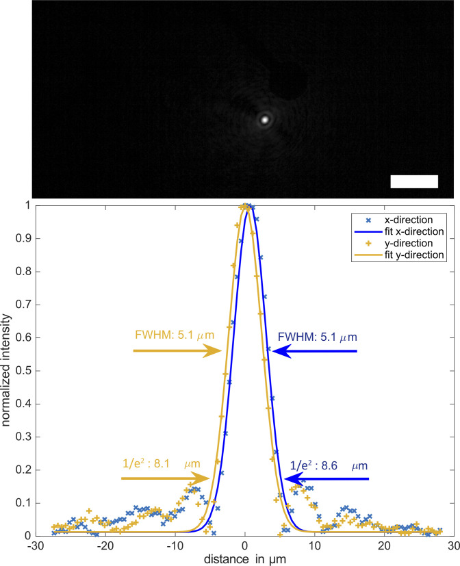
Figure S6. Example of a single focus observed through an multi-electrode array at 20× magnification with an inverted microscope.
Spatial resolution is defined by the width of a single focus. Gaussian fits to the data revealed focus widths of about 5-μm FWHM or 8-μm 1/e2 width along x and y direction of the camera grid. Scale bar is 50 μm.

