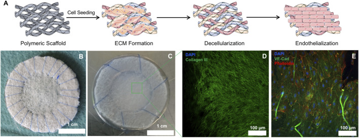
FIGURE 1.
Macroscopic and microscopic appearance of hTEMs before and after in vitro culture. (A): Schematic representation of the hTEM production procedure (Images adapted from Servier Medical Art under a creative common attribution 3.0 unported license). Briefly, PGA/P4HB scaffolds were sutured on stainless-steel metal ring and seeded with hDFs. After 4 weeks culture, patches were decellularized and finally endothelialized in vitro. (B): Macroscopic image of the PGA/P4HB scaffold sutured onto a stainless-steel metal ring before cell seeding. (C): Macroscopic appearance of the hTEM resulting from 4 weeks of tissue culture and subsequent decellularization. The hTEM is characterized by a shiny and smooth surface, indicating the presence of ECM. (D): Confocal microscope image of the hTEM stained for collagen III (green) and DAPI (blue) reveals the presence of a dense and homogeneous collagenous matrix with complete absence of cell nuclei. (E): Confocal microscope image showing homogeneous endothelialization on the hTEM surface.

