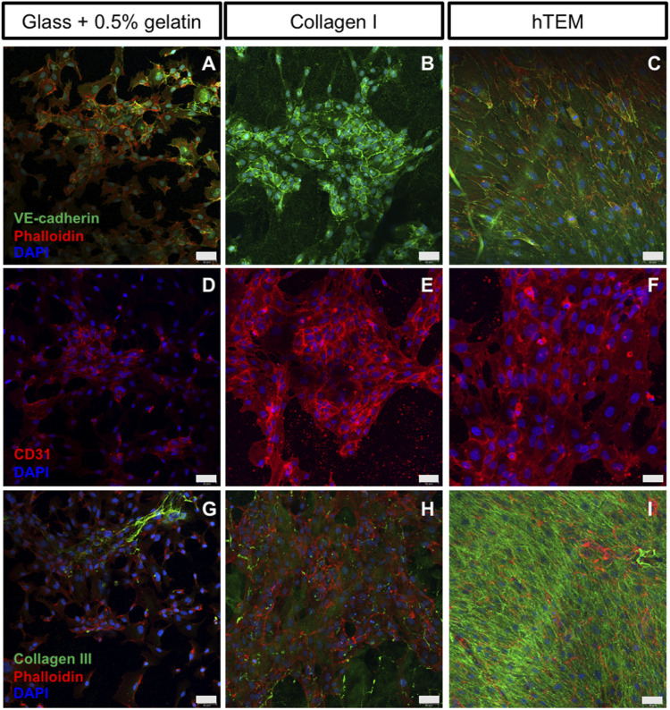
FIGURE 2.
Confocal images of ECFCs cultured on gelatin-coated glass, collagen I gels, and hTEMs at 5 days. (A–C): ECFCs stained for VE-cad (green), phalloidin (red), and DAPI (blue). (D–F): ECFCs stained for CD31 (red) and DAPI (blue). (G–I): Staining for Collagen III (green), phalloidin (red), and DAPI (blue). (A,D,G): glass covered with 0.5% gelatin show limited pre-endothelialization potential with poor ECFCs confluency after 5 days of culture, limited cell-cell contact, and low expression of CD31 and VE-cad. (B,E,H): Collagen I gel show spot-dependent pre-endothelialization. ECFCs are organized in colonies with presence of cell-cell contacts and expression of CD31 and VE-cad. (C,F,I): ECFCs seeded onto hTEM substrates demonstrate good pre-endothelialization potential, by achieving complete confluency after 5 days of culture (50 µm scale bars).

