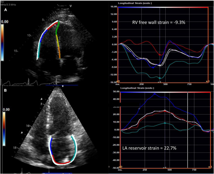
FIGURE 2.
Abnormal speckle tracking strain in diffuse cutaneous systemic sclerosis. (A) Example of reduced right ventricular free wall strain (normal <-20%) and (B) reduced left atrial reservoir strain (normal >39%) from the apical four chamber view in patients with systemic sclerosis. The white curve represents the average of the peak systolic strain curves. RV, right ventricle; LA, left atrium.

