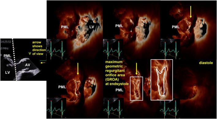
Figure 4.
Illustration of the dynamics of geometric regurgitant orifice area (GROA) in a patient with primary mitral regurgitation by 3D transesophageal echocardiography (TEE). On the left side the sectional plane of a TEE long axis view is shown. The dotted line represents the cutting plane of the en-face view to the GROA. The direction of view is illustrated by the yellow arrow. AV, aortic valve; LV, left ventricle; PML, posterior mitral leaflef. Right to this 2D image six consecutive 3D-en-face views to the GROA are presented illustrating the maximum GROA at end systole.

