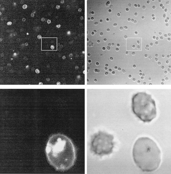
FIG. 5.
Fluorescence microscopy of a treated malaria sample. Parasite cultures at the young trophozoite stage were exposed to rhodaminated K4-S4(1–13)a (1 μM; 1 to 2 min; RT), washed twice in culture medium, and observed unfixed under a microscope. Images were taken within 5 min. The upper left picture is an optical section (rhodamine filter) of a field of treated RBC. The upper right picture is the light transmission of the same field. Each white rectangle defines the zone enlarged, shown in the corresponding lower pictures.

