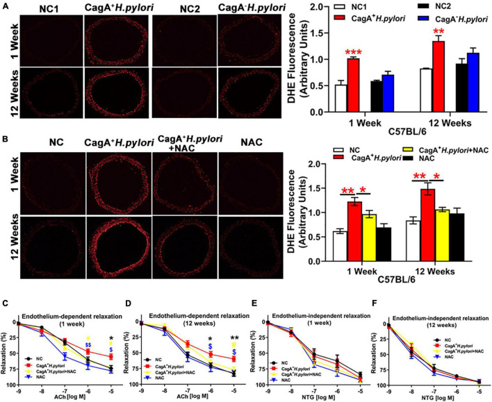
FIGURE 2.
CagA+ H. pylori, not CagA– H. pylori infection, impaired endothelial function through increased ROS production in mice. Representative fluorescent images and quantification of ROS formation in the aorta of male C57BL/6 mice (A) (**P < 0.01, ***P < 0.001 by t-test) with CagA+ H. pylori, CagA– H. pylori or PBS gavage. Aortic ROS production was significantly increased in mice with CagA+ H. pylori infection, not with CagA– H. pylori infection. Treatment with NAC prevented aortic ROS production (B) (*P < 0.05,**P < 0.01 by one-way ANOVA) and preserved ACh-induced aortic relaxation (C,D) in mice with 1 or 12 weeks of CagA+ H. pylori infection, without change in NTG-induced aortic relaxation (E,F). *P < 0.05, **P < 0.01 (compared with NC), #P < 0.05 (compared with CagA+ H. pylori + NAC); $P < 0.05, $$P < 0.01 (compared with NAC) by one-way ANOVA. NC(1/2): normal control. NAC: N-acetylcysteine; ACh: acetylcholine; NTG: nitroglycerin. Data are presented as mean ± SEM; N = 8–10 mice for each group at each time point.

