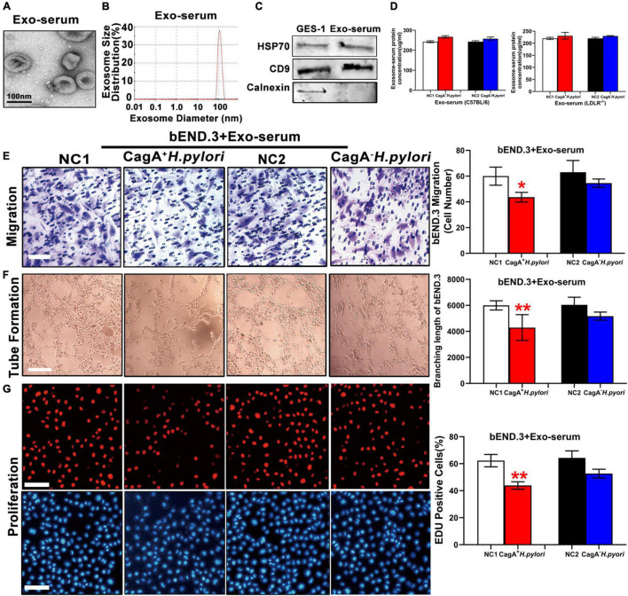
FIGURE 3.
Serum exosomes from CagA+ H. pylori infected mice impaired endothelial function. Mouse serum exosomes displayed typical features for exosomes including morphology on transmission electron microscopy (A) and size distribution (B). Western blotting analysis showed that the exosomes markers HSP70 and CD9 were present in the exosomes without the presence of calnexin (C). Although there was no significant difference in serum exosomes levels from mice infected with H. pylori compared to the controls (D), treatment with serum exosomes from mice with CagA+ H. pylori infection significantly inhibited the function of mouse bEND.3 cells in vitro with decreased migration (E, scale bars = 25 μm), tube formation (F, scale bars = 25 μm), and proliferation (G, scale bars = 100 μm). Exo, Exosomes; Exo-serum: exosomes isolated from mouse serum; NC(1/2): normal control. Data are resented as mean ± SEM; *P < 0.05; **P < 0.01 by t-test, N = 8–10 mice for each group. Experiment was repeated 3 times for every measurement.

