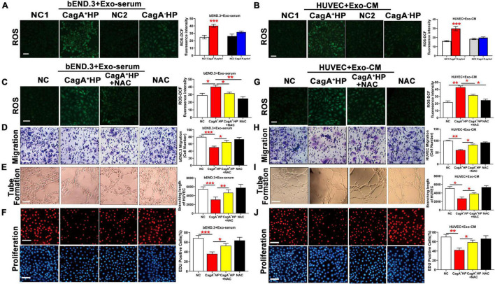
FIGURE 5.
CagA-containing exosome impaired endothelial function via increased ROS formation. (A) Representative fluorescent images and quantification of ROS formation in bEND.3 co-cultured with serum exosomes from mice infected with CagA+ H. pylori, CagA– H. pylori or PBS. Intracellular ROS formation was significantly increased in bEND.3 co-cultured with serum exosomes from mice infected with CagA+ H. pylori, not from mice infected with CagA– H. pylori or PBS. (B) Representative fluorescent images and quantification of ROS formation in HUVECs co-cultured with exosomes from conditioned media of GES-1 cultured with CagA+ H. pylori, CagA– H. pylori or PBS. Intracellular ROS formation was significantly increased in HUVECs co-cultured with exosomes from GES-1 cultured with CagA+ H. pylori, not from GES-1 co-cultured with CagA– H. pylori or PBS. NAC treatment significantly decreased intracellular ROS production and preserved endothelial function of bEND.3 (C–F) and HUVECs (G–J) treated with CagA-containing exosomes. NC(1/2): normal control; Exo-serum: exosomes from mouse serum; Exo-CM: Exosomes from conditioned medium; CagA+ HP: CagA+ H. pylori; CagA– HP: CagA– H. pylori.; NAC: N-acetylcysteine. Data were presented as mean ± SEM; *P < 0.05; **P < 0.01; ***P < 0.001 by t-test or one-way ANOVA. Experiment was repeated 3 times for every measurement. Scale bars (all, except for D,H,E) = 100 μm. Scale bars (D,H) = 25 μm. Scale bars (E) = 10 μm.

