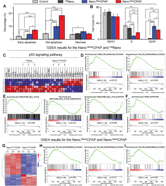
Figure 2.

Nano‐DimerCPAP potently activated p53 signaling cascades beyond Nano‐MonoCPAP in vitro. A) Apoptosis and necrosis analysis of NCI‐1650 cells incubated with PBS, Nano‐DimerCPAP (10 µg mL−1), Nano‐MonoCPAP (10 µg mL−1) or CtrlNano via flow cytometry for 48 h (n = 3, mean ± sd). B) Cell cycle analysis of NCI‐1650 cells treated with Nano‐DimerCPAP, Nano‐MonoCPAP, CtrlNano or PBS control for 48 h by FACS (n = 3, mean ± sd). C) Heat map of RNA‐Seq analysis of NCI‐H1650 cells’ mRNAs which were differentially expressed between Nano‐DimerCPAP and CtrlNano (n = 3). D) GSEA results for the p53 signaling pathway and the p53 downstream pathway. GSEA results for the REACTOME cell cycle checkpoints, E) the REACTOME cell cycle mitotic and F) the KEGG apoptosis. G) Hierarchical clustering of genes differentially expressed in NCI‐H1650 cells after exposure to Nano‐DimerCPAP for 24 h compared with Nano‐MonoCPAP (n = 3). H) GSEA analysis of Nano‐DimerCPAP versus Nano‐MonoCPAP showing the increased p53 signaling and downstream pathway. I) GSEA showing that the apoptosis of Nano‐DimerCPAP is superior to Nano‐MonoCPAP.
